Hip Instability
Anatomy of the Hip
The hip is a congruent ball and socket joint which is maintained by a series of ligamentous connections. The ball is the femoral head, which is the upper end of the femur (thigh bone). The deep socket is formed by the pelvis acetabulum, the part of the large pelvis bone that meets the upper end of the thigh bone.
Smooth, white tissue called articular cartilage covers the ends of the bones where they come together to form the hip joint. Cartilage acts as a cushion and allows the bones to glide over each other with very little friction. A strong fibrocartilage ring-like structure surrounding the socket (acetabulum) is called the hip labrum. This labrum articulates with the ball (femoral head) to create a suction seal that keeps the ball in the socket and provides stability to the hip joint.
There are four main ligaments of the hip joint. One ligament, the ligamentum teres, resides within the hip joint directly connecting the ball to the socket. The other three ligaments help stabilize the joint from the outside. Working together, these structures form a joint responsible for both bearing weight and range of motion.
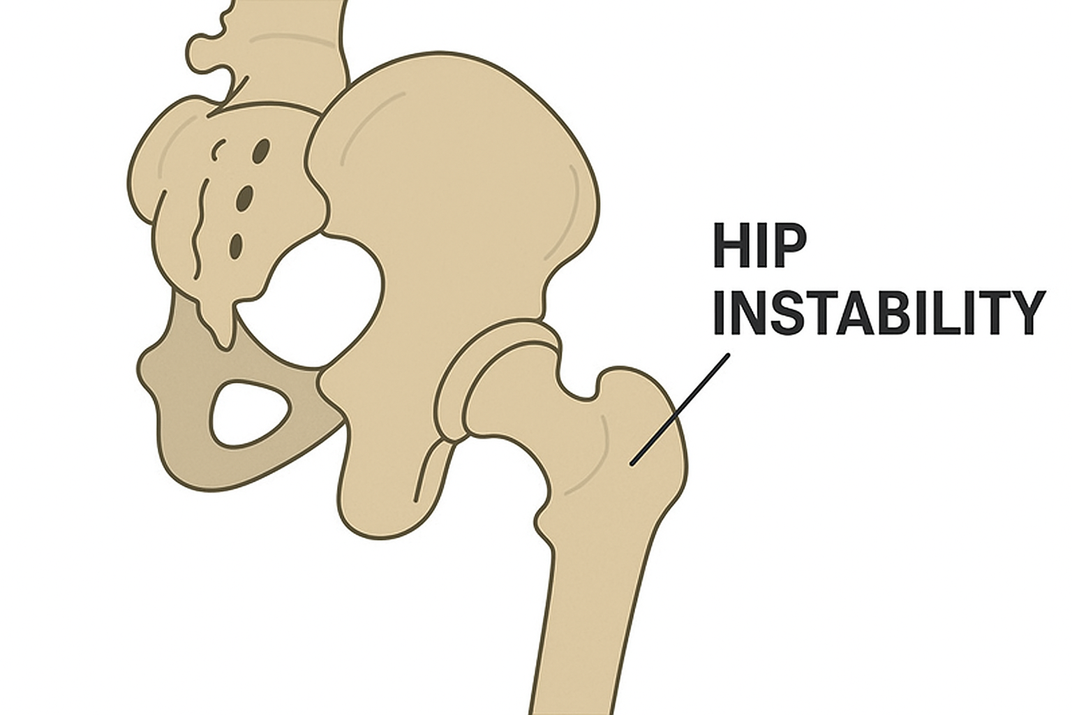
In normal hip anatomy, the femoral head is suction sealed within the acetabulum allowing for smooth joint movement. Hip instability is typically defined as unsteadiness of the hip joint and pathologic hip motion. The term can be used to cover several conditions where pain or weakness affect the normal function of the hip. Hip instability can be caused by an accident, sports injury, or a congenital condition.
Hip instability is most often divided into a traumatic or atraumatic classification. It is classically associated with developmental dysplasia of the hip, yet also commonly presents itself in people who participate or have previously participated in sports requiring large ranges of motion.
Traumatic instability
Traumatic hip injury can be caused by a major force, such as a motor vehicle crash, a fall from a significant height, or a collision while playing a sport. A traumatic injury can damage the femoral head, acetabulum, articular cartilage, labrum, or ligaments. Since hip stability is dependent on the relationship between these bony and soft tissues structures, damage to these structures can cause hip instability.
Atraumatic hip instability
Atraumatic injury can be caused by anatomic abnormalities of the hip or by repetitive motion in professional athletes. In athletes, hip instability may begin with subtle anatomic abnormalities that are exacerbated by repetitive hip joint rotation and axial loading. Atraumatic hip instability can result from numerous causes, including the following medical conditions:
- Hip dysplasia: When the bones of the hip socket are formed with a less than normal amount of femoral head coverage, the hip joint can wear out faster and become partially or completely dislocated. Most people with hip dysplasia are born with the condition, and infants are typically assessed for hip dysplasia shortly after birth. Hip instability in infancy is referred to as developmental dysplasia of the hip (DDH). In milder cases, the condition may not cause symptoms until early adulthood. When diagnosed in adolescence, hip dysplasia is called acetabular dysplasia.
- Joint hyperlaxity: The hip has several ligaments connecting the femur to the pelvis. Laxity of the four ligaments which stabilize the hip can result in pain and hip instability. Hypermobility, or extreme flexibility, can be a result of trauma or inherited connective tissue disorders.
- Femoroacetabular impingement (FAI): FAI is a condition where extra bone grows along either the socket (acetabulum) or ball (head of femur) forming the hip joint. This causes the femur and acetabulum to rub against each other during ranges of motion such as sitting, kneeling, squatting, jumping, or running. Eventually, the friction damages the cartilage or labrum that cushions the hip, leading to hip instability, limited range of motion, and pain.
The most common symptoms of hip instability are:
- Hip or groin pain
- Movement beyond the normal range of motion (laxity)
- A sensation of the hip coming out of the socket
- A clicking, snapping, or popping sound in the hip
- Subluxation (partial dislocation) or dislocation
Hip instability can be difficult to diagnose because it often occurs without any clear signs or symptoms. However, some people may experience pain, clicking, or popping in the hip joint. In addition, hip instability can lead to a feeling of looseness in the joint or a sensation that the joint is giving way.
In many patients with atraumatic hip instability, there may not be a clear etiology. As a result, the experts at LALL Orthopedics + will diagnose hip instability based on a complete patient history, comprehensive physical examination, and radiographic imaging findings.
Most atraumatic cases can be successfully managed with conservative, non-surgical treatments such as physical therapy, anti-inflammatory medications, taping, therapeutic ultrasound, healing biologic interventions (platelet-rich plasma or stem-cell injections), shockwave therapy and activity modification. Minimally invasive, same-day surgery to repair injured structures such as the labrum or tighten the lax (stretchy) ligaments may be indicated should symptoms remain following a course of conservative measures.
The most common surgical treatment for hip instability is a procedure called arthroscopic stabilization. This surgery is performed by making small incisions in the hip joint and inserting a tiny camera, called an arthroscope. The surgeon then uses special instruments to repair or tighten the muscles, ligaments, and tendons around the hip joint. In some cases, the surgeon may also need to remove damaged tissue or bone.
Another surgical option for treating hip instability is a procedure called periacetabular osteotomy (PAO). This surgery is performed by making an incision in the hip joint and repositioning the socket of the hip bone. This helps to stabilize the joint and improve the function of the hip.
Recovery from hip instability surgery usually takes several weeks. During this time, patients will need to use crutches or a cane to help them walk. Most patients will be able to return to their normal activities within four to six months.
Dr. Ajay C. Lall is a former dual sport NCAA collegiate athlete (football and track & field), American board certified and triple fellowship-trained hip surgeon. Dr. Lall and the team at LALL Orthopedics + specializes in diagnosing and treating hip instability. Schedule a hip consultation today.
LALL Orthopedics + has offices in Paramus, NJ, Philadelphia, PA and Belvidere, IL. Our team regularly sees patients from Bergen County, Hackensack, and Morristown, NJ.
At a Glance
Ajay C. Lall, MD, MS, FAAOS
- Board Certified – Orthopedic Surgery
- Triple Fellowship Trained
- Performs over 750 Surgeries Per Year
- Learn more